
- Home
-
प्रमुख समाचार
 Bhupender Yadav, chaired the 91st Meeting of the Standing Committee of the National Board for Wildlife
Pashu Sandesh, 09 July 2026 Union Minister for…
 Punjab Govt Launched hi tech Cold Van for Veterinary Biologicals
Pashu Sandesh,08 July 2026 To strengthen the safe…
 उत्तर प्रदेश : पशुधन बीमा प्रीमियम पर 85% अनुदान देगी सरकार
Pashu Sandesh, 07 July 2026 उत्तर प्रदेश…
 Godrej Bets Big on India’s Booming Pet Food Market, Expands ‘Ninja’ Brand Across South India
Pashu Sandesh, 07 July 2026 In a…
 International Tiger Week at Ranthambore Signals a New Era in India's Tiger Conservation Journey
Pashu Sandesh, 05 July 2026 As conservationists,…
 India's National One Health Mission Conducts 5-Day Zoonotic Outbreak Mock Drill 'Pashujanya Yudh Abhyas'
Pashu Sandesh, 03 July 2026 The Department…
-
बड़े मुद्वे
 Victory for UP Vets as Government agrees to table NPA proposal within one month in the cabinet meet
Pashu Sandesh, 04 October 2017
-
एक्सक्लूसिव रिपोर्ट
 Veterinary Associations of India- Himachal Pradesh Veterinary Officers Association (HPVOA)
Pashu Sandesh, 26 October 2017
-
डेयरी
-
कैंपस
-
पोल्ट्री
-
वाइल्ड लाइफ
 The valedictory ceremony of the Wildlife week organised at National Zoological Park Delhi
Pashu Sandesh, 8 October 2021
 राष्ट्रीय प्राणी उद्यान, दिल्ली में आयोजित 67वें वन्यजीव सप्ताह का समापन समारोह
Pashu Sandesh, 8 October 2021
-
एनिमल वैलफेयर
 उ.प्र. गौशाला स्वावलंबन अभियान:मुख्यमंत्री योगी ने प्रयास की सराहना
Pashu Sandesh, 29 जनवरी 2026 डॉक्टर आर बी…
 पशु मित्र संपादकीय टीम में चार समर्पित पशु प्रेमियों की नियुक्ति, संपादकीय बोर्ड अन्य प्रतिभाओं की खोज में : पशु मित्र पत्रिका
Pashu Sandesh, 24 jan 2026 देश की जानी-मानी…
 पशु प्रेमियों द्वारा शहर में पशु टीकाकरण कार्यक्रम में चारु चौधरी ने महिला प्रकोष्ठ के लिए अनुपमा द्विवेदी के नाम घोषित किया-इस अवसर पर एडबलूबीआई,…
Pashu Sandesh, 02 July 2025 गोरखपुर (उत्तर प्रदेश):…
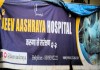 लखनऊ शहर में बेसहारा पशुओं के लिए 'जीव आश्रय'एक आशा की किरण,संस्था ने गत वर्ष 141,536 से अधिक जानवरों को बचाया
Pashu Sandesh, 05 April 2025 डॉ. आर.बी. चौधरी…
 समस्त महाजन जल्द ही 2 एम्बुलेंस के साथ 24 घंटे पशु बचाव सेवा शुरू करने जा रहा है - राष्ट्रीय राजधानी में अपनी तरह की…
Pashu Sandesh, 20 December 2024 “यदि सभी पशु…
 गोबर- गोमूत्र से गांव की अर्थव्यवस्था सुधारने मैं जुटी एक स्वयंसेवी संस्था समस्त महाजन के उपलब्धियां पर महान हस्तियों की नजर
Pashu Sandesh, 10 Sep 2024 Dr R B…
-
रोजगार
-
राज्यों की खबरें
-
पेट केयर
 Why Does Leptospira Persist In The Kidneys Of Carrier Animals?
Pashu Sandesh,04 September 2017 Parvinder Kaur Lubana…
-
किसान कैफ़े
 अदरक और हल्दी की खरीद और विपणन के लिए समझौता ज्ञापन पर हस्ताक्षर
उत्तर प्रदेश के किसानों की टीम "रूरल हब"…
-
आई सी ए आर
- महत्वपूर्ण लिंक


Persistent or patent urachus in calf
Pashu Sandesh, 13 March 2020
VAIBHAV BHARDWAJ*, GAURAV KUMAR
Introduction:
Persistent or patent urachus is a congenital condition in which embryonic connection (called urachus) between the urinary bladder and allantoic sac of a fetus fails to close after birth causing dribbling of urine from the umbilical region of the newborn. In simple words, we can say that the urachus is a long tube connecting the urinary bladder and belly button of the fetus, which usually closed after birth. But if it fails to close after birth, a condition called persistent or patent urachus. This condition can be simple or sometimes associated with omphalitis, congenital stricture or occlusion of the urethra. Newborn affecting with this condition show continuous dribbling of urine from umbilical region days after birth and the surrounding area remains wet.
Causes of patent urachus:
Congenital aetiology of patent urachus is unknown but may be presumed due to torsion of the umbilical cord during fetal life or sometimes may occur due to tension on umbilicus during parturition causing dilation of the urachus which prevents their occlusion or incomplete involution. It may occur as an acquired condition associated with umbilical infection/inflammation or any cause of increased intra-abdominal pressure due to meconium retention (atresia ani).
Clinical signs:
- Free flow urination or dribbling of urine through the umbilical opening.
- A hard tube or cord-like structure caudal to the umbilicus is palpated.
- Mildly distended abdomen.
- Area surrounding umbilicus remains wet.
Surgical treatment:
The calf is placed on dorsal recumbency and the ventral abdomen is prepared antiseptically. Local analgesia (2% Lignocaine hydrochloride) is infiltered into the surgical site. A skin incision is made around the umbilicus and extended to the abdominal wall. Traction is applied to the umbilicus and the urachus is traced up to the bladder and is ligated by chromic catgut as close to the bladder as possible. The surgical area is digitally palpated to check for the presence of adhesions. The skin is closed with non-absorbable braided silk material using interrupted horizontal mattress.
Postoperative management:
- Broad-spectrum antibiotics and analgesic should be administered intramuscularly for 5-7 days.
- Prevention of operated site from soil, faeces or urine contamination.
- Antiseptic cleaning of the surgical wound and insect repellent spray 3-4 times a day up to recovery.
- Skin sutures removal after 7-10 days.
- In the case of pus or maggots at the surgical site, report the veterinarian immediately.
VAIBHAV BHARDWAJ*, GAURAV KUMAR
Department of Veterinary Surgery and Radiology, College of Veterinary Sciences, Lala Lajpat Rai University of Veterinary and Animal Sciences, Hisar, Haryana, India.


