
- Home
-
प्रमुख समाचार
 Aspergillosis in Poultry: The Hidden Mold Behind Heavy Losses
Pashu Sandesh, 31 March 2026 Km Himani Assistant…
 Yogi Adityanath Emphasizes Self-Sustaining Gaushalas: Shares Views in Recent Meeting
Pashu Sandesh, 29 Jan 2026 Dr RB Chaudhary
 Pashu Mitra Welcomes New Editorial Team Members: Invites More Talent
Pashu Sandesh, 24 Jan 2026 Dr Rb choudhary
 Effect of Heat Stress on Growth Performance and Serum Metabolites in Broilers and the Protective Role of Phytogenic Formulations
Pashu Sandesh, 21 Jan 2026 Dr.…
 Guidelines and SOPs for Blood Transfusion and Blood Banks for Animals released by GOI
Pashu Sandesh, 25 August 2025 …
-
बड़े मुद्वे
 Victory for UP Vets as Government agrees to table NPA proposal within one month in the cabinet meet
Pashu Sandesh, 04 October 2017
-
एक्सक्लूसिव रिपोर्ट
 Veterinary Associations of India- Himachal Pradesh Veterinary Officers Association (HPVOA)
Pashu Sandesh, 26 October 2017
-
डेयरी
-
कैंपस
-
पोल्ट्री
-
वाइल्ड लाइफ
 The valedictory ceremony of the Wildlife week organised at National Zoological Park Delhi
Pashu Sandesh, 8 October 2021
 राष्ट्रीय प्राणी उद्यान, दिल्ली में आयोजित 67वें वन्यजीव सप्ताह का समापन समारोह
Pashu Sandesh, 8 October 2021
-
एनिमल वैलफेयर
 उ.प्र. गौशाला स्वावलंबन अभियान:मुख्यमंत्री योगी ने प्रयास की सराहना
Pashu Sandesh, 29 जनवरी 2026 डॉक्टर आर बी…
 पशु मित्र संपादकीय टीम में चार समर्पित पशु प्रेमियों की नियुक्ति, संपादकीय बोर्ड अन्य प्रतिभाओं की खोज में : पशु मित्र पत्रिका
Pashu Sandesh, 24 jan 2026 देश की जानी-मानी…
 पशु प्रेमियों द्वारा शहर में पशु टीकाकरण कार्यक्रम में चारु चौधरी ने महिला प्रकोष्ठ के लिए अनुपमा द्विवेदी के नाम घोषित किया-इस अवसर पर एडबलूबीआई,…
Pashu Sandesh, 02 July 2025 गोरखपुर (उत्तर प्रदेश):…
 लखनऊ शहर में बेसहारा पशुओं के लिए 'जीव आश्रय'एक आशा की किरण,संस्था ने गत वर्ष 141,536 से अधिक जानवरों को बचाया
Pashu Sandesh, 05 April 2025 डॉ. आर.बी. चौधरी…
 समस्त महाजन जल्द ही 2 एम्बुलेंस के साथ 24 घंटे पशु बचाव सेवा शुरू करने जा रहा है - राष्ट्रीय राजधानी में अपनी तरह की…
Pashu Sandesh, 20 December 2024 “यदि सभी पशु…
 गोबर- गोमूत्र से गांव की अर्थव्यवस्था सुधारने मैं जुटी एक स्वयंसेवी संस्था समस्त महाजन के उपलब्धियां पर महान हस्तियों की नजर
Pashu Sandesh, 10 Sep 2024 Dr R B…
-
रोजगार
-
राज्यों की खबरें
-
पेट केयर
 Why Does Leptospira Persist In The Kidneys Of Carrier Animals?
Pashu Sandesh,04 September 2017 Parvinder Kaur Lubana…
-
किसान कैफ़े
 अदरक और हल्दी की खरीद और विपणन के लिए समझौता ज्ञापन पर हस्ताक्षर
उत्तर प्रदेश के किसानों की टीम "रूरल हब"…
-
आई सी ए आर
- महत्वपूर्ण लिंक


DIAPHRAGMATIC HERNIA
Pashu Sandesh, 18 October 2023
Ankit Dangi1and Devika Dangi2
1. Assistant Professor, Veterinary Surgery and Radiology, International Institute of Veterinary Education & Research, Rohtak
2. Animal Nutrition, Lala Lajpat Rai University of Veterinary & Animal Sciences, Hisar
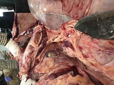
Diaphragmatic hernia (DH) is a thoracic-digestive disorder in which there is the evisceration of abdominal viscera into the thoracic cavity through a congenital or acquired defect in the diaphragm. The continuity of the diaphragm is disrupted such that abdominal organs can migrate into the thoracic cavity.
Abdominal organs that herniate:
- Most commonly- Reticulum
- May get involved – omasum, abomasums, loops of intestine, spleen or liver
It causes chronic ruminal tympany, anorexia and displacement of the heart. It is a chronic wasting and inflammatory disorder in adult buffaloes and cows, and also in buffalo bulls. In small animals mostly it is caused by trauma.
Aetiology:
- Weakening of the diaphragm by the lesions of TRP.
- Congenital weak points of the diaphragm.
- Increased abdominal pressure during pregnancy or parturition.
- Foreign body – reticular contraction – pricking of the diaphragm constantly – perforation.
- Areas at the junction of muscular and tendinous portion – more prone – lack of tone and thickness.
- Trauma
Buffaloes are more prone than cattle:
- The right ventromedial tendinous zone is much thinner
- Pericardiophrenic vessels missing
- Lesser collagen content
Clinical signs:
- Recurrent tympany – proportional to the portion of reticulum herniated – more severe adhesions.
- Aspiratory pneumonia – regurgitation – adhesions cause distortion and derangement in normal alignment of oesophageal groove, cardia and reticulo-omasal opening – sojourn of ingesta in forestomach.
- Abrupt fall in milk yield.
- Scant defecation or diarrhoea with a foul smell.
- Brisket oedema with or without jugular pulsation.
- Abduction of elbow.
- Chronic cough rarely.
- If untreated – inanition – progressive emaciation
– dehydration – death.
- Suspended rumination.
Diagnosis:
- Clinical signs
- Auscultation – muffled cardiac sounds, reticular sounds cranial to 6th rib.
- Roentgenograms/Radiography:(contrast or simple)
- Right lateral recumbency – most hernia in right hemidiaphragm.
- Supine position – lateral projection – empty reticulum appears as an air-filled viscous in the thoracic cavity.
- Exploratory rumenotomy
- Ultrasonography
Treatment:
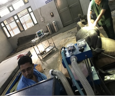
Firstly:
Laprorumenotomy
- Complete evacuation of rumen
- Removal of any foreign body if present
- Add prebiotic and probiotic
Secondly :
Herniorrhaphy
- Ventral midline abdominal post-xiphoid approach
- Off-feed for 48 hours following rumen evacuation
- i/v fluids during fasting (polyionic solutions with dextrose)
- Regular monitoring- ECG, pulse rate, SpO2, blood pressure, heart rate.
- Negative pressure within the chest cavity is created by suction of air under a water seal
- Extensive adhesions with the pulmonary lobe require partial or complete lobectomy
- Postoperative considerations
- Antibiotics for 5 – 7 days
- Analgesic for 3 - 5 days.
- Fluid therapy
- Easily digestible feed
- Pregnant animal – check for viable foetus
- Suture ASD and removal after 10 – 12 days
- Consider vet advice regularly


