
- Home
-
प्रमुख समाचार
 Yogi Adityanath Emphasizes Self-Sustaining Gaushalas: Shares Views in Recent Meeting
Pashu Sandesh, 29 Jan 2026 Dr RB Chaudhary
 Pashu Mitra Welcomes New Editorial Team Members: Invites More Talent
Pashu Sandesh, 24 Jan 2026 Dr Rb choudhary
 Effect of Heat Stress on Growth Performance and Serum Metabolites in Broilers and the Protective Role of Phytogenic Formulations
Pashu Sandesh, 21 Jan 2026 Dr.…
 Guidelines and SOPs for Blood Transfusion and Blood Banks for Animals released by GOI
Pashu Sandesh, 25 August 2025 …
-
बड़े मुद्वे
 Victory for UP Vets as Government agrees to table NPA proposal within one month in the cabinet meet
Pashu Sandesh, 04 October 2017
-
एक्सक्लूसिव रिपोर्ट
 Veterinary Associations of India- Himachal Pradesh Veterinary Officers Association (HPVOA)
Pashu Sandesh, 26 October 2017
-
डेयरी
-
कैंपस
-
पोल्ट्री
-
वाइल्ड लाइफ
 The valedictory ceremony of the Wildlife week organised at National Zoological Park Delhi
Pashu Sandesh, 8 October 2021
 राष्ट्रीय प्राणी उद्यान, दिल्ली में आयोजित 67वें वन्यजीव सप्ताह का समापन समारोह
Pashu Sandesh, 8 October 2021
-
एनिमल वैलफेयर
 उ.प्र. गौशाला स्वावलंबन अभियान:मुख्यमंत्री योगी ने प्रयास की सराहना
Pashu Sandesh, 29 जनवरी 2026 डॉक्टर आर बी…
 पशु मित्र संपादकीय टीम में चार समर्पित पशु प्रेमियों की नियुक्ति, संपादकीय बोर्ड अन्य प्रतिभाओं की खोज में : पशु मित्र पत्रिका
Pashu Sandesh, 24 jan 2026 देश की जानी-मानी…
 पशु प्रेमियों द्वारा शहर में पशु टीकाकरण कार्यक्रम में चारु चौधरी ने महिला प्रकोष्ठ के लिए अनुपमा द्विवेदी के नाम घोषित किया-इस अवसर पर एडबलूबीआई,…
Pashu Sandesh, 02 July 2025 गोरखपुर (उत्तर प्रदेश):…
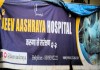 लखनऊ शहर में बेसहारा पशुओं के लिए 'जीव आश्रय'एक आशा की किरण,संस्था ने गत वर्ष 141,536 से अधिक जानवरों को बचाया
Pashu Sandesh, 05 April 2025 डॉ. आर.बी. चौधरी…
 समस्त महाजन जल्द ही 2 एम्बुलेंस के साथ 24 घंटे पशु बचाव सेवा शुरू करने जा रहा है - राष्ट्रीय राजधानी में अपनी तरह की…
Pashu Sandesh, 20 December 2024 “यदि सभी पशु…
 गोबर- गोमूत्र से गांव की अर्थव्यवस्था सुधारने मैं जुटी एक स्वयंसेवी संस्था समस्त महाजन के उपलब्धियां पर महान हस्तियों की नजर
Pashu Sandesh, 10 Sep 2024 Dr R B…
-
रोजगार
-
राज्यों की खबरें
-
पेट केयर
 Why Does Leptospira Persist In The Kidneys Of Carrier Animals?
Pashu Sandesh,04 September 2017 Parvinder Kaur Lubana…
-
किसान कैफ़े
 अदरक और हल्दी की खरीद और विपणन के लिए समझौता ज्ञापन पर हस्ताक्षर
उत्तर प्रदेश के किसानों की टीम "रूरल हब"…
-
आई सी ए आर
- महत्वपूर्ण लिंक


Important Rickettsial Diseases of animals
Pashu Sandesh, 19th July 2018
Sonali Mishra, Kunal Pandit, Gaytri Kashyap
RICKETTSIAL DISEASES
These are Gram negative, non-spore forming, non-motile, small obligate intracellular highly pleomorphic bacteria. Their cell wall is bigger than virus but smaller than bacteria. They have DNA and RNA and also have an ATP transport system that allows them to use host ATP, so replication within the cytoplasm of eukaryotic host cells. Arthropod acts as reservoirs and vectors (e.g., ticks, mites, lice or fleas) for the disease. These are sensitive to antibiotics.
Need for studying animal Rickettsioses-
- Economic losses to livestock industries
- Mental and emotional distress to the owners
- Financial costs to owners- via veterinary treatments, tick-prevention programs
BOVINE ANAPLASMOSIS/ GALLSICKNESS
- Anaplasma phagocytophilum – Zoonotic
- A. marginale - Clinical outbreaks
- A. centrale - limited pathogenicity
Prevalence- All six continents, especially in tropical areas
Physical Vectors-
Flies, Horse Flies, Stable Flies, Dear Flies, Mosquitoes
Instruments
Surgical Instruments, Dehorning Instruments, Castration Instruments, Hypodermic Needles
Biological Vector
Ticks
PATHOGENESIS
- RBCs Phagocytosis
- Anemia
- Lack of O2& Nutrients in tissues
- Animal remains carrier
- Calves Under 6 Months- Show no Signs or Symptoms
- Up to 18 months old- Show only Symptoms
GROSS LESIONS
- Severe anemia with pale mucous membrane and icterus.
- Spleen is greatly enlarged and reddish in colour.
- Liver and the gall bladder is distended with granular bile.
- Petechiae of epicardium and catarrhal gastroenteritis are evident.
- Liver is also enlarged with rounded edges.
MICROSCOPICAL LESIONS
- Hyperplasia of bone marrow.
- Extramedullary haematopoiesis in spleen and liver is observed.
DIFFERENTIAL DIAGNOSIS
Other haemolytic diseases- Babesia, Post parturient bovine haemoglobinuria
Heart water disease
Causative agent: Ehrlichia ruminantium
Animal Transmission
Vector-borne – Amblyomma ticks.
Larvae and nymphs infected from infected animals.
Clinical Signs
Incubation period: 14 to 28 days
Four forms of disease
I. Peracute (rare)
II. Acute (most common)
III. Subacute (rare)
IV. Mild or subclinical
Acute form:Most common form
- Sudden fever (107oF)
- Inappetence, depression
- Tachypnea, respiratory distress
- Nervous signs
V. Chewing movements, eyelid twitching, tongue protrusion, circling, paddling
- Death within 1 week
Post Mortem Lesions
Hydropericardium, Hydrothorax, Ascites, Pulmonary & mediastinal edema, Lymphadenopathy, Congestion and edema in the brain.
MICROSCOPIC LESIONS- Edematous and generalized luecocytic infiltration in tissues, demonstration of organism in vascular endothelium.
Diagnosis:
- Clinical signs
Fever, respiratory distress, sudden death
- Presence of Amblyomma ticks
- Laboratory Tests
- Microscopic identification organism
- PCR, IFA, ELISA
Canine Monocytic Ehrlichiosis (Tracker dog disease, Canine haemorrhagic fever, Tropical canine pancytopaenia)
Causative agent- Ehrlichia canis
Vector:The Brown Dog Tick (Rhipicephalus sanguineus) usually spreads Ehrlichia canis
Clinical signs
Nonspecific signs –
Fever, Anorexia & lethargy, lymphadenopathy, splenomegaly & weight loss, vomiting, diarrhea, lameness, edema in the hind legs, dyspnea & purulent oculo-nasal discharge.
Bleeding disorders - Anemia, mild epistaxis, petechiae and ecchymoses, death - Consequence of hemorrhages or secondary infections.
Ocular signs:
Anterior uveitis, corneal opacity and tortuous retinal vessels, corneal opacity, bleeding haemorrhages on gums.
POSTMORTEM LEISONS
Hemorrhage in mucosa of GI tract, urogenital tract, kidneys, edematous and haemorrhagic lymph node, and edema of limbs.
MICROSCOPIC LESIONS
Perivascular accumulation of RE cells, plasma cells in meninges, liver, kidney and lymph reticular organs, bone marrow hypo plastic, degeneration and centrilobular necrosis in liver
Diagnosis
Dog with fever, enlarged lymph nodes, bleeding, or arthritis in multiple joints.Low platelet numbers, high globulin levels, and mild anemia on blood testing, canine thrombocytopaenia, pancytopenia. Ehrlichia canis seen in membrane-bound inclusions (morulae) within the cytoplasm of a monocyte (buffy coat smear, Wright stain).
Sonali Mishra1, Kunal Pandit2, Gaytri Kashyap1
1Division of Veterinary Pathology, ICAR-Indian Veterinary Research Institute, Izatnagar – 243 122, Bareilly, U.P, India
2Department of Veterinary Anatomy, C.V.A.Sc., GBPUAT, Pantnagar – 263 145, Uttarakhand, India


