
- Home
-
प्रमुख समाचार
 Aspergillosis in Poultry: The Hidden Mold Behind Heavy Losses
Pashu Sandesh, 31 March 2026 Km Himani Assistant…
 Yogi Adityanath Emphasizes Self-Sustaining Gaushalas: Shares Views in Recent Meeting
Pashu Sandesh, 29 Jan 2026 Dr RB Chaudhary
 Pashu Mitra Welcomes New Editorial Team Members: Invites More Talent
Pashu Sandesh, 24 Jan 2026 Dr Rb choudhary
 Effect of Heat Stress on Growth Performance and Serum Metabolites in Broilers and the Protective Role of Phytogenic Formulations
Pashu Sandesh, 21 Jan 2026 Dr.…
 Guidelines and SOPs for Blood Transfusion and Blood Banks for Animals released by GOI
Pashu Sandesh, 25 August 2025 …
-
बड़े मुद्वे
 Victory for UP Vets as Government agrees to table NPA proposal within one month in the cabinet meet
Pashu Sandesh, 04 October 2017
-
एक्सक्लूसिव रिपोर्ट
 Veterinary Associations of India- Himachal Pradesh Veterinary Officers Association (HPVOA)
Pashu Sandesh, 26 October 2017
-
डेयरी
-
कैंपस
-
पोल्ट्री
-
वाइल्ड लाइफ
 The valedictory ceremony of the Wildlife week organised at National Zoological Park Delhi
Pashu Sandesh, 8 October 2021
 राष्ट्रीय प्राणी उद्यान, दिल्ली में आयोजित 67वें वन्यजीव सप्ताह का समापन समारोह
Pashu Sandesh, 8 October 2021
-
एनिमल वैलफेयर
 उ.प्र. गौशाला स्वावलंबन अभियान:मुख्यमंत्री योगी ने प्रयास की सराहना
Pashu Sandesh, 29 जनवरी 2026 डॉक्टर आर बी…
 पशु मित्र संपादकीय टीम में चार समर्पित पशु प्रेमियों की नियुक्ति, संपादकीय बोर्ड अन्य प्रतिभाओं की खोज में : पशु मित्र पत्रिका
Pashu Sandesh, 24 jan 2026 देश की जानी-मानी…
 पशु प्रेमियों द्वारा शहर में पशु टीकाकरण कार्यक्रम में चारु चौधरी ने महिला प्रकोष्ठ के लिए अनुपमा द्विवेदी के नाम घोषित किया-इस अवसर पर एडबलूबीआई,…
Pashu Sandesh, 02 July 2025 गोरखपुर (उत्तर प्रदेश):…
 लखनऊ शहर में बेसहारा पशुओं के लिए 'जीव आश्रय'एक आशा की किरण,संस्था ने गत वर्ष 141,536 से अधिक जानवरों को बचाया
Pashu Sandesh, 05 April 2025 डॉ. आर.बी. चौधरी…
 समस्त महाजन जल्द ही 2 एम्बुलेंस के साथ 24 घंटे पशु बचाव सेवा शुरू करने जा रहा है - राष्ट्रीय राजधानी में अपनी तरह की…
Pashu Sandesh, 20 December 2024 “यदि सभी पशु…
 गोबर- गोमूत्र से गांव की अर्थव्यवस्था सुधारने मैं जुटी एक स्वयंसेवी संस्था समस्त महाजन के उपलब्धियां पर महान हस्तियों की नजर
Pashu Sandesh, 10 Sep 2024 Dr R B…
-
रोजगार
-
राज्यों की खबरें
-
पेट केयर
 Why Does Leptospira Persist In The Kidneys Of Carrier Animals?
Pashu Sandesh,04 September 2017 Parvinder Kaur Lubana…
-
किसान कैफ़े
 अदरक और हल्दी की खरीद और विपणन के लिए समझौता ज्ञापन पर हस्ताक्षर
उत्तर प्रदेश के किसानों की टीम "रूरल हब"…
-
आई सी ए आर
- महत्वपूर्ण लिंक


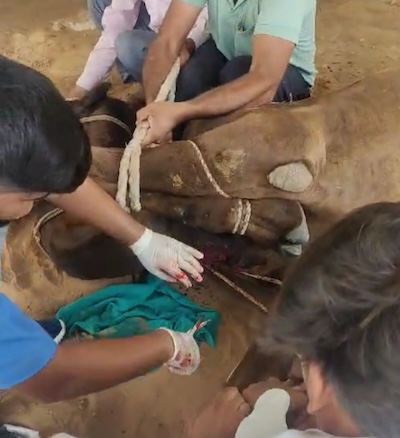
Surgical management of fibroma in a camel (camelus dromedarius)
Pashu Sandesh, 06 November 2023
Yogendra Singh
Department of Surgery and Radiology, RPS Veterinary College, Balana, Mahendergarah – 123029, Haryana, India
Fibroma is a noncancerous (benign) tumour or growth of fibrous, connective tissue. They can occur almost anywhere in and on your body. The majority of the tumours are round to oval intradermal or subcutaneous masses. The present report deals with successful surgical management and histo-pathological diagnosis of fibroma in a dromedary camel.
History and Clinical Examination
A 5-year-old male camel (Camelus dromedarius) of about 500 Kg body weight was presented to (TVCC) Teaching Veterinary Clinical Complex, RPS Veterinary Hospital, Balana, Mahendergarh with a history of large hard swelling at brisket region for 15 months (Fig. 1). Owner reported animal decreases feed and water intake since one month. The camel was initially examined for evaluation of the hard swelling. The owner also reported that the animal was medicinally treated by a local veterinarian. The swelling was increased in size with time.
There was ulceration on the brisket of the swelling and pus discharge was noticed. A maggot infestation was also reported by the owner that was treated successfully and bleeding had also been reported occasionally. History and clinical findings suggested it to be a fibroma. At the time of the checkup at TVCC, the general checkup respiratory rate, temperature and pulse rate of the animal were normal range. The animal felt some pain and discomfort while touching the brisket site.
Diagnosis-
Fibromas are diagnosed by history, physical examination, symptoms and medical history. Depending on the type of growth. To confirm the diagnosis these tests may include:
Treatment
Feed and water were withheld for 24 hours prior to surgery. The camel was restrained in sternal recumbency and sedated with xylazine, 150 mg intravenously. After securing it in the right lateral recumbency the surgical site was aseptically prepared using soap and water and scrubbed. About 15 ml of 2% lignocaine hydrochloride was infiltrated locally at the surgical site before the surgical procedure. An elliptical skin incision was made around the base of the hard swelling. Following a blunt dissection the blood vessels were ligated using chromic catgut no 2. Complete enucleation of tumour mass weighing 2.5 kg was done (Fig. 2). The skin incision was closed with a horizontal mattress suture pattern using silk no 2. Postoperatively, the wounds were irrigated with povidone iodine solution on alternate days and lorexane ointment was applied topically. Wounds were covered with a sterile gauge followed by a multi-tail bandage. Broad spectrum antibiotic (Dicrysticin 5 gm) I.M. for 5 days and analgesic (Phenylbutazone 3000 mg) I.M. for 3 days were given parenterally. The sutures were removed on the 15th postoperative day. The open wound healed in 20 days.
A gross examination of the excised tumour revealed it to be hard. Histopathological examination revealed cells with hyperchromatic nuclei spindle-shaped with an abundance of collagenous fibres. The cells were uniform but arranged in a dysplastic manner. The proliferating blood vessels were present in abundance (Fig 3).
Discussion
Treatment of a tumour or growth presents a serious challenge. Position and function lead to continuous soiling and irritation of the wound. To prevent this, a multi-tail bandage was applied. At this moment the tumour or growth no longer comes in contact with the soil when the animal lies down. Mesenchymal tumours of the skin and soft tissues comprise a wide range of entities, some of which are of uncertain classification. Fibromas are benign neoplasms of fibrocytes with abundant collagenous stroma which has a rare occurrence in large animals. They often occur in adults, sex and species do not affect its prevalence. The majority of tumours are round to oval intradermal or subcutaneous masses. The secondary bacterial infection was controlled with broad-spectrum antimicrobial therapy and dressing with povidone-iodine solution in this case. However, the chest pad wounds need protection from getting soiled during the healing period as this practice prevents contamination


