
- Home
-
प्रमुख समाचार
 निराश्रित गौवंश पालने पर मिलेंगे 6000 रुपए प्रति माह
मुख्यमंत्री निराश्रित/बेसहारा गोवंश सहभागिता योजना Pashu Sandesh, 08…
 India Unveils Its First WOAH‑Certified Equine Disease Free Compartment at RVC Meerut
Pashu Sandesh, 05 July 2025 In a historic…
 President Murmu Proclaims Animals as 'Jeevan Dhan' at 11th IVRI Convocation
Pashu Sandesh, 30th june 2025
 गोरखपुर के पशु प्रेमियों की एकदिवसीय कार्यशाला संपन्न
उत्तर प्रदेश महिला आयोग की उपाध्यक्ष चारु चौधरी…
 भारत में चारा प्रणालियों को मजबूत करना: प्रभावी प्रबंधन और खाद्य सुरक्षा के लिए एक खाका
Pashu Sandesh, 09 June 2025
-
बड़े मुद्वे
 Victory for UP Vets as Government agrees to table NPA proposal within one month in the cabinet meet
Pashu Sandesh, 04 October 2017
-
एक्सक्लूसिव रिपोर्ट
 Veterinary Associations of India- Himachal Pradesh Veterinary Officers Association (HPVOA)
Pashu Sandesh, 26 October 2017
-
डेयरी
-
कैंपस
-
पोल्ट्री
-
वाइल्ड लाइफ
 The valedictory ceremony of the Wildlife week organised at National Zoological Park Delhi
Pashu Sandesh, 8 October 2021
 राष्ट्रीय प्राणी उद्यान, दिल्ली में आयोजित 67वें वन्यजीव सप्ताह का समापन समारोह
Pashu Sandesh, 8 October 2021
-
एनिमल वैलफेयर
 पशु प्रेमियों द्वारा शहर में पशु टीकाकरण कार्यक्रम में चारु चौधरी ने महिला प्रकोष्ठ के लिए अनुपमा द्विवेदी के नाम घोषित किया-इस अवसर पर एडबलूबीआई,…
Pashu Sandesh, 02 July 2025 गोरखपुर (उत्तर प्रदेश):…
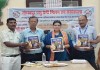
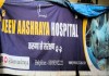 लखनऊ शहर में बेसहारा पशुओं के लिए 'जीव आश्रय'एक आशा की किरण,संस्था ने गत वर्ष 141,536 से अधिक जानवरों को बचाया
Pashu Sandesh, 05 April 2025 डॉ. आर.बी. चौधरी…
 समस्त महाजन जल्द ही 2 एम्बुलेंस के साथ 24 घंटे पशु बचाव सेवा शुरू करने जा रहा है - राष्ट्रीय राजधानी में अपनी तरह की…
Pashu Sandesh, 20 December 2024 “यदि सभी पशु…
 गोबर- गोमूत्र से गांव की अर्थव्यवस्था सुधारने मैं जुटी एक स्वयंसेवी संस्था समस्त महाजन के उपलब्धियां पर महान हस्तियों की नजर
Pashu Sandesh, 10 Sep 2024 Dr R B…
 गोवंश को राष्ट्रीय पशु घोषित कराने के लिए राष्ट्रव्यापी अभियान
Pashu Sandesh, 27 November 2022 ओसियां…
-
रोजगार
-
राज्यों की खबरें
-
पेट केयर
 Why Does Leptospira Persist In The Kidneys Of Carrier Animals?
Pashu Sandesh,04 September 2017 Parvinder Kaur Lubana…
-
किसान कैफ़े
 अदरक और हल्दी की खरीद और विपणन के लिए समझौता ज्ञापन पर हस्ताक्षर
उत्तर प्रदेश के किसानों की टीम "रूरल हब"…
-
आई सी ए आर
- महत्वपूर्ण लिंक


Surra: Oxidative stress determines a disease's prognosis
Pashu Sandesh, 06 January 2022
Dr Vivek Agrawal*, Dr. Mukesh Shakya, Dr. A.K. Jayraw, Dr. Nidhi Singh, Dr G.P. Jatav, and Dr. Nirmala Jamra
- Introduction
Surra, or animal trypanosomosis, is a tropical and subtropical disease caused by Trypanosoma evansi (a eukaryotic, unicellular, euryxenous, haemoflagellate organism). Due to the disease's detrimental effect on animal productivity, it has a significant societal and economic impact. T. evansi infection is transmitted mechanically by a variety of haematophagous biting flies and affects a wide variety of domestic and wild animals. In India, animal trypanosomosis is a negative input into the conversion of resources into products, goods, and services. As a result of the disease's catastrophic effect on animal health and productivity, considerable economic losses arise. In Africa, animal trypanosomosis costs around US$5 billion per year, with direct losses ranging between US$1 and US$1.2 billion and an annual cost of approximately $30 million for disease control, which includes curative and preventative approaches.
- Oxidative stress
In Trypanosome infection, oxidative stress occurs when there is an imbalance between reactive oxygen generation and the inability to readily detoxify reactive intermediates or repair the ensuing damage. It is critical to maintaining a reducing environment, which is maintained by enzymes by a constant input of metabolic energy. Disruptions in this natural redox state can have a harmful effect by producing peroxides and free radicals, which can cause damage to all cell components, including proteins and lipids.
Superoxide (O 2), hydrogen peroxide (H2O2), peroxyl radicals (ROO), and hydroxyl radicals (OH) are all highly reactive unpaired electron or free radicals. This is produced during T. evansi infections, resulting in the formation of oxidative products and tissue damage. Macrophages appear to be a critical target cell for parasite action in African trypanosomiasis because activated macrophages from mouse models of infection create free radicals such as nitric oxide (NO), oxygen (O 2), and O 2 -derived H2O2. These free radicals are generated when activated macrophages respond to parasite antigens with pro-inflammatory cytokines such as tumour necrosis factor-alpha (TNF-a), gamma interferon (IFN-c), interleukin 1 beta (IL-1B), and IL-6. NO appears to have been produced during parasite infection of T. evansi by reacting with O 2 to form peroxynitrite anion (ONOO) or by the Fenton reaction to form OH. Both ONOO and OH are the most active oxidising agents, initiating lipid peroxidation primarily through interactions with polyunsaturated fatty acids in cell membranes and exhibiting necrotic consequences. The syndromes associated with this parasite infection are serious, and numerous medicines have been utilised to treat the sickness produced by T. evansi. Nitric oxide (NO) can also enter the erythrocyte membrane and combine with sulfhydryl (SH) groups, forming nitrosyl-haemoglobin. In the presence of oxygen, this progressively oxidises to methemoglobin (MetHb). From a functional aspect, oxidative damage to the plasma membrane affects its fluidity and permeability, increasing cell fragility as a result. Free radicals have been reported to act as cytotoxic agents Due to their function as an O2 and CO2 transporter, the erythrocyte is constantly exposed to free radicals. However, red blood cells possess a strong antioxidant defence that converts free radicals to significantly less reactive intermediates. Apart from their potential impacts on the peroxidation of membrane phospholipids, free radicals can also damage membrane proteins directly, resulting in negative functional effects.
- Lipid peroxidation
Lipids, particularly polyunsaturated fatty acids, are susceptible to oxidation by free radicals, giving rise to the term lipid peroxidation, which is characterised by the presence of malondialdehyde (MDA). MDA accumulation in tissues or biological fluids is indicative of the degree to which free radicals are generated, oxidative stress is induced, and tissue damage occurs. The findings indicate that T. evansi infection is related to oxidative stress, as evidenced by lipid peroxidation, and that these oxidative alterations impair erythrocytes' ability to resist oxidative damage to the erythrocyte membrane. Oxidative stress and alterations in antioxidant enzymes have also been observed in rats infected with T. evansi, as evidenced by an increase in glucose-6-phosphate dehydrogenase (G6P-DH) and glutathione peroxidase. The increased activity of G6P-DH during T. evansi infections is due to the production of reactive oxygen species (ROS) by erythrocytes, which promotes lipid peroxidation and activates the host cell's hexose monophosphate shunt. Peroxidation of lipids has a number of detrimental effects, including increased membrane stiffness, impaired receptor function, and altered permeability.
- Conclusion
In T. evansi infection, oxidative stress occurs when "insulted" erythrocytes release reactive oxygen species (ROS), which promotes lipid peroxidation. The increased erythrocytic oxidation and decrease in haematological indices suggest that increased erythrocytic oxidation is involved in erythrocytic destruction and progression of anaemia in T. evansi infection.
Corresponding Author:
Dr Vivek Agrawal
Associate Professor Department of Veterinary Parasitology
Dr Mukesh Shakya, Assistant Professor, Department of Veterinary Parasitology
Dr A.K. Jayraw, Professor and Head, Department of Veterinary Parasitology
Dr Nidhi Singh, Assistant Professor, Department of Veterinary Medicine
Dr G.P. Jatav, Associate Professor, Department of Veterinary Pathology
Dr Nirmala Jamara, Assistant Professor, Department of Veterinary Parasitology
College of Veterinary Sciences & A.H
Nanaji Deshmukh Veterinary Science University
Mhow, Indore -453446, Madhya Pradesh(India)


